Cultured shrimps suffer from various diseases due to infectious and non-infectious causes. Infectious diseases are caused by viruses, bacteria, fungi and certain parasites. Treatment cannot be carried out effectively when shrimp diseases occur in a pond. The best way to get rid of diseases is by practicing good farm management or prevention. In this regard, information on various kinds of diseases and their prevention procedures are useful.
Infectious Diseases
(i) Monodon Baculovirus Disease (MBV)
Etiological Agent
MBV-type or PmSNPV is a type A occluded monodon baculovirus.
Clinical Signs
Lethargy, anorexia, poor feeding, dark colouration and reduced growth rate. Infected shrimps are often associated with fouling of gills and appendages by ciliates such as Zoothamnium spp. and Vorticella spp. Acute infection leads to loss of epithelial cells of hepatopancreas.
Treatment
No treatment available for MBV infection
Prevention and Control
There is little information on prevention and control of the MBV infection in shrimp pond culture. The prevention method for the MBV infection is possibly through avoidance by screening the PL's before stocking shrimp in the pond.
(ii) Hepatopancreatic Parvo-like Virus (HPV) Disease
Etiological Agent
HPV is caused by a small parvo-like virus, 22-24 nm in diameter.
Clinical Signs
Reduced feeding, poor growth rate, body surface and gill fouling with ciliates and occassional opacity of abdominal muscles. Severe infections may include a whitish and atrophied hepatopancrease, anorexia and reduced preening activity. Losses may be occur due to the increased occurance of surface and gill fouling organisms and secondary infections by the opportunistic Vibrio spp.
Treatment
No treatment available for HPV infection.
Prevention and Control
No information is available on the prevention and control procedures for HPV infection. However, screening the PLs before stocking shrimp by routine histology or the Giemsa-impression smear method is recommended.
(iii) Yellow-head Disease (YHD)
Etiological Agent
Yellow-headed virus (YHV) is a ssRNA, rod shaped, enveloped virus with two rounded ends.
Clinical Signs
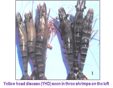
The affected shrimp shows a marked reduction in food consumption. Following this, a few moribund shrimp will appear swimming slowly near the surface of the pond dike and remain motionless. The animals have pale bodies, a swollen cephalothorax with a light yellow to yellowish hepatopancreas and gills. A high mortality rate may reach 100% of affected populations within 3-5 days from the onset of disease.
Treatment
No treatment is available for YHV infection.
Prevention and Control
The reliable method to prevent the occurrence of YHD is possibly through avoidance, such as careful selection of post larvae, reduction or elimination of horizontal transmission including carriers, disinfection of contaminated ponds or equipment with 30 ppm; and chlorine, providing shrimp with good waterquality and proper nutrition.
(iv) White Spot Disease (WSD)
Etiological Agent
The disease is caused by the dsDNA virus, Systemic Ectodermal and Mesodernal Baculovirus (SEMBV).
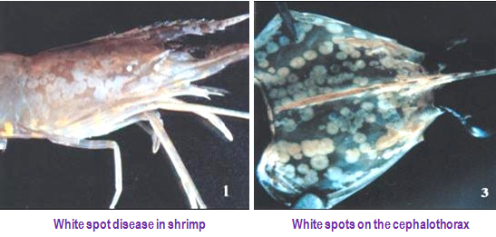
Clinical Signs: Clinically affected shrimp were first seen to swim to the water surface and congregate at the pond dikes. Typical clinical signs include white spots or patches, 1-2 mm in diameter, on the inside of the shell and carapace, accompanied by reddish discoloration of the body. SEMBV is able to cause acute epizootics of 5-10 days duration with mortality rate from 40% to 100%.
Clinical Signs: Clinically affected shrimp were first seen to swim to the water surface and congregate at the pond dikes. Typical clinical signs include white spots or patches, 1-2 mm in diameter, on the inside of the shell and carapace, accompanied by reddish discoloration of the body. SEMBV is able to cause acute epizootics of 5-10 days duration with mortality rate from 40% to 100%.
Diagnosis Procedure
The diagnosis procedure of SEMBV infection is based on the appearance of the intranuclear hypertrophy in stained histological sections and the presence of virus particles in the nucleus of the infected cells observed under the electron microscope. PCR technique is recently used to detect SEMBV in shrimp larval and other stages, including broodstock and subclinical virus carriers.
Treatment
No treatment is available for SEMBV infection.
Prevention and Control
Prevention practices through avoidance are strongly recommended for the farmers, involving the combinations of efficient pond management, use of proper feed, selection of good quality of PL, reduction of possible carriers, avoidance of introduction of contaminated water into the pond, and disinfection of all equipment and utensils.
(Source: FAO technical paper)
(v) Infectious Hepatopancreatic and Lymphoid Organ Necrosis (IHLN)
Etiological Agent
The primary cause of the disease is attributed to viral etiology.
Clinical Signs
Light pinkish to yellowish discolouration of the cephalothorax region. Often fouling by ciliate protozoan Zoothamnium seen. Blackened and necrotic hepatopancreas. Secondary bacterial infection from bacteria such as Vibrio alginolyticus seen.
Treatment
No treatment is available for IHLN infection.
Prevention and Control
Keep the physico-chemical condition of pond environment within acceptable levels. To avoid bacterial and viral pathogen entering from outside, closed culture could be useful in prevention of IHLN disease.
(i) Luminous Vibriosis
Etiological Agent
Vibrio harveyi, Vibrio vulnificus
Clinical Signs
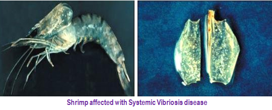
High mortality rate in young juvenile shrimp (one month syndrome). Moribund shrimp hypoxic often come to the pond surface and edges of pond. Vertical swimming behavior immediately before onset of acute mortality. Presence of luminescent shrimp in ponds.
Treatment
Disinfection of intake water with Formalin (100-200 ppm). Administration of Oxolinic acid (0.6 ppm) and Sarafloxacin (5mg/kg) through feed for 5 days.
Prevention and Control
Proper pond and water management. Utilization of reservoir for intake water.
(ii) Vibriosis
Etiological Agent
Vibrio vulnificus, V. parahemolyticus, V. alginolyticus, V. anguillarum, V. damsella, V. fluvialis and V. mimicus.
Clinical Signs
High mortality rates, particularly in young juvenile shrimp. Moribund shrimp with corkscrew swimming behavior appear at edge of pond. Reddish discoloration of juvenile shrimp.
External Fouling
Black spots, chronic soft shelling.
Treatment
Disinfection of intake water i.e. formalin 100-200 ppm. Anti-microbial preparation application through feeds (Oxolinic acid 0.6 ppm and Sarafloxacin 5 mg/kg).
Prevention and Control
Proper pond and water management and utilization of reservoir for intake water.
(i) Larval Mycosis
Etiological Agent
Filamentous fungi of genus Lagenidium spp. and other filamentous fungi, such as Sirolpidium spp. and Haliphthoros spp.
Clinical Signs
Eggs and larve are weak and appear whitish. Moratlities may reach 100% within two days. Fungal mycelium replaces the larval tissues and ramifies into all parts of the body and protrudes out of the body and develops into sporangia.
Prevention and Control
General hatchery management practices such as use of UV sterilised and filtered seawater, adequate water exchange etc., must be strictly followed. Rearing water, equipment used in the hatchery and all hatchery facilities must be thoroughly disinfected before retarting the hatchery operations.
(i) Black Gill Disease
Etiological Agent
Fusarium spp
Clinical Signs
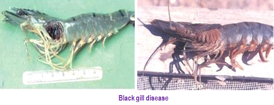
Brownish to blackish discoloration on the gills of juvenile shrimp.
Treatment
No treatment is available for fungal infestation without harming the shrimp.
Prevention and Control
No information on prevention and control. However, good management of the pond bottom and prevention of the entry of wild crustaceans into the pond, which may carry pathogen, can be effective control practices.
(ii) Surface Fouling Diseases
Etiological Agent
Many species of bacteria, algae and protozoa such as filamentous bacteria, Leucouthrix sp., Flavobacterium sp. and Zoothamnium sp.
Clinical Signs
Infected shrimps show black/ brown gills or appendage discoloration or fuzzy/cottony appearance due to a heavy colony of the organisms. In some cases, the severely affected shrimp die during the molting period.
Treatment
Chlorine and formalin are often used to treat those commensal organisms if shrimp display heavy infection. Changing water is the most preferable management, which stimulates molting of the shrimp in order to reduce the infestation.
Prevention and Control
Prevention and control of the occurrence of surface fouling are usually done through maintenance of good sanitary conditions at the pond bottom and the overall pond area. Organic matters and suspended solids in the pond should be reduced to prevent the attachment of those fouling organisms. This is achieved by changing the water or applying lime.
(iii) Microsporidosis (Cotton shrimp disease or Milk shrimp disease)
Etiological Agent
Microsporidia such as Thelohania spp., Nosema spp., and Pleistophora spp.
Clinical Signs
Infected shrimps appear opaque and cooked. Gradual and low levels of mortalities are observed. Microsporidia invade and replace gill, muscle, heart, gonads and hepatopancreas, and cause necrosis in these regions.
Prevention and Control
Maintain of good sanitary conditions at the pond bottom and the overall pond area.
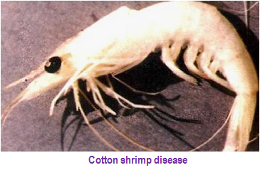
(Source: CIBA Extension Series)
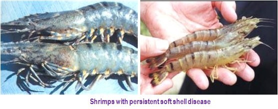
(i) Soft-shell syndrome
Etiological Agent
The exact cause of soft-shell syndrome is not known. However, low saline condition in the culture pond and deterioration of pond bottom condition are some physico-chemical factors that causes this disease. Shrimps fed with low protein diet, contamination through agricultural run-off, high soil pH, low water phosphate and low organic matter in soil all have an impact on soft-shell disease.
Clinical Signs
Shrimps are weak, usually off-feed, have a loose thin exoskeleton. Rostrum is stiff as healthy shrimps. Wavy undulating intestine is clearly visible.
Prevention and Control
Low stocking density, feeding with high quality feed and frequent water exchange are likely to reduce the recurrence of the disease.
No comments:
Post a Comment